La spécificité du tadalafil est liée à sa longue demi-vie, permettant une action qui excède largement celle des autres inhibiteurs de PDE5. L’absorption digestive est complète, avec un pic plasmatique atteint en 2 heures environ. Le métabolisme est réalisé via CYP3A4, produisant des métabolites inactifs éliminés principalement dans les fèces. La sélectivité enzymatique est élevée, réduisant les effets indésirables extra-caverneux. Les réactions indésirables fréquentes incluent céphalées, bouffées vasomotrices et troubles digestifs légers. L’activité pharmacologique est stable, indépendamment de l’ingestion d’aliments. Dans les comparaisons de longue durée, acheter cialis pas cher est mentionné en relation avec les études portant sur la persistance d’efficacité et la constance de la cinétique plasmatique.
Njgonline.nl
Geologie en Mijnbouw / Netherlands Journal of Geosciences 79 (1): 59-71 (2000)
Composition and genesis of rattlestones from Dutch soils as shown by Mössbauer spectroscopy, INAA and XRD J.J. van Loef 1
Interfacultair Reactor Instituut, TU Delft , Mekelweg 15, 2629 JB DELFT, The Netherlands;
Manuscript received: 1999; accepted in revised form: 29-12-1999
Abstract
The chemical and mineralogical composition of rattlestones found near the main Dutch rivers has been studied by Möss-bauer spectroscopy, INAA and XRD. Rattlestones are concretions of iron, formed in an environment of lateral iron accumu-lation, under the influence of periodical oxidation, around a fine core of ferruginous sediments, mainly clay and sand. Thecore has shrunk and detached itself from the mantle around it. 57Fe Mössbauer spectroscopy was applied to identify the ironoxides, among which goethite is predominant. The goethite crystallinity was investigated by measuring its magnetic propertiesand its crystallinity, which is poorest at the outer side of the stone. The latter is confirmed by the broadening of the different X-ray reflections. In addition, illite and vermiculite were identified by XRD; these clay minerals were found mainly in the core.
The elemental composition was determined by INAA. The iron content in the mantle is about 50% by weight and gradual-
ly decreases outwards, while the core contains 2-15% Fe by weight. Differences between rattlestones from the Middle Pleis-tocene East of the Meuse river and those from the Late Pleistocene North of it are the absence of lepidocrocite and a richermineralogy in the former.
It is concluded that the rattlestones are formed around a fine clayey core. Groundwater supplied the iron and other (trace)
elements for the genesis. It is unlikely that rattlestones are the result of oxidation of siderite. Keywords: crystallinity, goethite, iron accumulation, lepidocrocite, Mössbauer spectroscopy, rattlestones, siderite, trace elements
Introduction
soluble layers have been removed by solution, leavinga central part detached from the outer part, such as a
Rattlestones have been known for long. They were re-
concretion of iron oxide filled with loose sand that
ferred to as ‘aetites’ or ‘eagle stones’ in Roman times
rattles on being shaken. Van der Burg (1969) suggest-
(Adams, 1938; Bromehead, 1947). In spite of this,
ed that rattlestones are formed by oxidation of side-
surprisingly little information is available on the
rite concretions which were deposited contemporane-
chemical and mineralogical composition of these
ously with the sediments in the beds where they are
stones. Few previous publications dealt with rattle-
stones (Van der Burg, 1969) and with the climatolo-
The geochemical and mineralogical characteristics
gical factors limiting their distribution in the Dutch
of the rattlestones were investigated for the present
Pleistocene (Van der Burg, 1971). The present contri-
bution discusses the composition of several rattle-stones found near the main rivers in the Netherlands,
Material
According to a recent definition (Jackson, 1997), a
Eleven rattlestones have been investigated. They were
rattlestone is a concretion composed of concentric
collected in several sand and gravel pits near the main
laminae of different composition, in which the more
Dutch rivers, i.e. at Koningsbosch East of the Meuse
Geologie en Mijnbouw / Netherlands Journal of Geosciences 79(1) 2000
river (Sterksel Formation, three specimens), and
(Coney, 1988). The structural order of iron oxides en-
North of the Meuse at Deest, Lathum and Wapenveld
countered in natural environments ranges from rea-
(Kreftenheye Formation, four specimens) and at
sonably good to seemingly amorphous. This has a
Leersum and Schaarsbergen (Drenthe Formation,
great influence on the magnetic properties. Devia-
four specimens). The codes used for these sites are
tions of the magnetic properties of iron oxides of very
Ko, De, La, Wa, Le and S, respectively; the Roman
small particle size from those of coarse-grained coun-
numbers I and II are used wherever necessary to dis-
terparts lead to radical changes in their Mössbauer
tinguish between stones from one single site. An alter-
spectra (Van der Kraan & Van Loef, 1966; Murad,
ated siderite concretion found in the Reuver clay at
1996). This is illustrated by the hyperfine field, which
about 30 m depth (Kiezeloöliet Formation) was stud-
has a single value at any temperature in a pure mag-
netic crystal, but can vary greatly or even disappear at
The specific density of the stones was obtained by
room temperature in a soil iron oxide like goethite.
determining volume and weight. The latter varied be-
This type of spectroscopy can be used to distinguish
tween 30 and 500 g. The rattlestones were opened
between the two valence states of iron, e.g. in goethite
carefully in order to collect the loose, yellowish fine
grains of the core, all of which weighed only a few
Mössbauer spectroscopy is based on the recoil-free
percent of the stone. The specific density of the outer
resonance absorption of gamma rays in certain atom-
part of the stone (called ‘mantle’), which was always
ic nuclei, for example the stable iron isotope 57Fe (2%
less than 10 mm thick, was obtained and the size of
natural abundance). Gamma rays with an energy of
14.4 keV are emitted by radioactive 57Co with a half-
Material from the core and several concentric lami-
life of 270 days and the resonant absorption cross
nae of the mantle of each rattlestone were powdered
section of these (γ rays in 57Fe is very high at room
separately. The lamination is irregular and the lami-
temperature. The method allows determination of nu-
nae with a width of 1-2 mm frequently merge into one
clear energy levels to an extremely high accuracy, so
another (Van der Burg, 1969). Differences in hardness
that slight variations caused by different interactions
and color are helpful in making a separation between
between electrons and the nucleus become measur-
laminae. The interior material of the mantle is hardest
able. These interactions reflect differences in the elec-
and often also the darkest; the exterior material of the
tronic, magnetic, geometric or defect structure in
rattlestone is mostly quite soft and easy to scrape off
solids. The spectroscopy is based upon the principle
the mantle. In total about 60 samples from the various
that, by moving the radioactive source, a very small
rattlestones were investigated. Core samples are coded
energy shift can be attained in the emitted gamma
1 and samples from the interior and exterior mantle
material have been coded 2 and 3; in case more layers
The instrumental setup consists of the 57Co-source,
in the mantle are distinguishable, the consecutive lay-
detector and the iron-containing sample in between;
ers have been numbered 2, 3 and 4. Material from dif-
in such a transmission experiment, the Mössbauer
ferent shells of the siderite concretion was sampled
spectrum (MS) obtained consists of the intensity of
and numbered 1 (central segment), 2 (first shell), 3
the (γ rays measured in the detector, plotted as a
(second one) and so on. In practice about 200 mg ma-
function of the source velocity. A minimum in MS
terial per sample sufficed for detailed analysis. Mun-
corresponds with resonance absorption in the sample
sell hues were used to determine the color of each
under investigation. A major value of Mössbauer
sample (Cornell & Schwertmann, 1996).
spectroscopy as an analytical tool lies in the fact thatany iron contained in a solid must show up in MS, in-
dependent of sample crystallinity or, of the form inwhich Fe is bound (Murad, 1988). Sample prepara-
The methods used for the investigation are nuclear
tion is usually very simple; the sample thickness is
techniques: Mössbauer spectroscopy (Kuzmann et
al.,1998) and instrumental neutron-activation analy-
The distinction by Mössbauer spectroscopy of
sis (INAA), which were complemented by X-ray dif-
goethite, hematite, lepidocrocite and siderite is based
on the magnetic hyperfine interaction. The first twooxides are magnetically ordered at room temperature
and MS of both goethite and hematite consist of six-line spectra. The hyperfine field, B, in goethite at
Mössbauer spectroscopy is a very useful technique for
room temperature is known to be 38.5 T and that in
investigating properties of Fe in soil iron oxides
hematite 51.5 T; the latter is used to calibrate the
Geologie en Mijnbouw / Netherlands Journal of Geosciences 79(1) 2000
source velocity in Mössbauer spectroscopy. TheMössbauer spectrum at 295 K (coded as MS[295K])of paramagnetic lepidocrocite consists of two lines ofequal intensity, the quadrupole splitting. The doubletis much larger in MS of siderite and readily distin-guished from that of lepidocrocite. Speciation in asample containing a few percent of Fe by weight isfeasible. Poorly crystalline iron oxides can still beidentified, in particular at lower temperatures. Spec-tral intensities of iron-containing species give infor-mation on their relative content in a sample. Furtherdetails on Mössbauer spectroscopy can be found inKuzmann (1988). Instrumental neutron-activation analysis
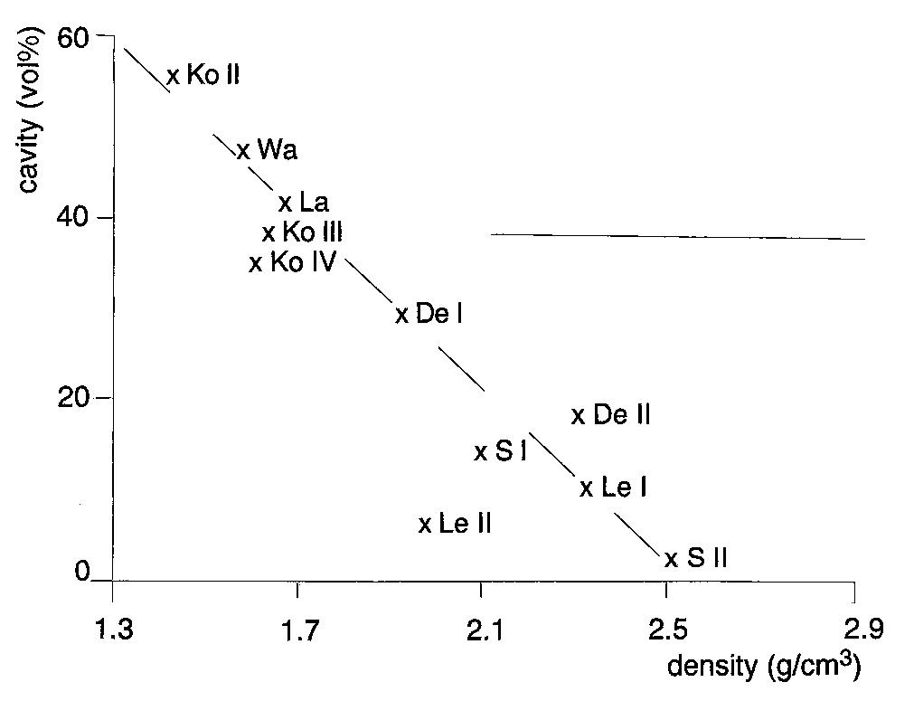
Fig.1. Cavity in the various rattlestones versus the density. The
dashed line is drawn as a guide to the eye. The horizontal line indi-
INAA is a quantitative method of high efficiency for
cates the range of densities of the stones investigated by Van der
the determination of a number of major and trace ele-ments and supplies geochemical information on thematerial that may be involved in the formation and
INAA has been applied at the nuclear reactor of
MS[295K] and MS[77K] of three samples from Ko
IRI at Delft to determine major elements, in particu-
II are shown in Figure 2. A central doublet dominates
lar Fe, and many trace elements. The standard devia-
the spectra at 295 K and has practically disappeared
tion and detection limit for Fe are less than 2% and
at 77 K. A six-line magnetic hyperfine splitting attrib-
about 100 ppm, respectively (Parry, 1991).
uted to goethite dominates MS[77K]. The doublet inMS[295K] is mainly due to goethite and to a limited
extent to clay minerals. This follows from their rela-tive intensities measured in MS[77K]. Remnants of a
XRD was performed to identify clay minerals. XRD
hyperfine splitting are still visible in the spectra of Ko
patterns were obtained that enabled to determine
II-1 and Ko II–2 at 295 K, but not in Ko II-3. The
peak positions and line widths of the iron oxides.
doublet resembles that of a paramagnet, but it is also
Diffractograms of about forty samples were made
characteristic of superparamagnetic goethite, due to
at the Laboratory of Soil Science and Geology of Wa-
extremely small crystallites. This is most pronounced
geningen University. The XRD was performed with a
in Ko II-3. In general, the sextets at 77K consist of
Philips PW 1710 diffractometer equipped with a
asymmetrically broadened lines indicating a distribu-
graphite monochromator using CoKα radiation.
tion of hyperfine fields due to a spread in goethite
Peaks were recorded from 5o to 75o 2Θ. The clay min-
crystallite size. The smaller the size, the lower the hy-
erals were thus identified, and the peak position and
perfine field (Van der Kraan, 1972). The distribution
linewidth of the goethite reflections were determined.
is narrowest in MS[77K] of Ko II-1 and widest inthat of Ko II-3, while the hyperfine field is highest in
the former and lowest in the latter (Table 1). Hence,the goethite crystallite size in the core is larger than in
The cavities of the rattlestones vary widely and can
take more than 50% by volume; this has a great effect
In MS[77K] of Ko II-1 and Ko II–3, the resonant
on the specific density of the rattlestone as a whole, as
absorption in the centre is attributed to non-magnetic
shown in Figure 1. The horizontal bar indicates the
minerals. It can be assigned to illite (XRD). Besides, a
density range of the numerous stones investigated by
small signal is measured at a source velocity of about
Van der Burg (1969), which only partly overlap with
3 mm/s that originates from an Fe2+ contribution; the
accompanying line of the doublet is submerged in the
The other characteristics, as shown by MS, INAA,
central part of the spectrum. X-ray diffractograms
XRD, and those regarding the hue of the specimens,
confirm the assignments of goethite and illite. In the
are dealt with in the following subsections.
XRD of Ko II-1, illite is more pronounced than in KoII-3 (see Table 2), which is contrary to the relative in-
Geologie en Mijnbouw / Netherlands Journal of Geosciences 79(1) 2000
Fig. 2. Mössbauer spectra of samples Ko II-1 (core), Ko II-2 and Ko II–3 (both mantle) at 77 and 295 K. A central doublet dominates
MS[295K] that has practically disappeared in MS[77K], where six-line spectra attributed to goethite are most pronounced. The positions of
the lines that correspond to the hyperfine field, B’, are indicated. A central doublet in MS[77K] of Ko II-1 and Ko II–3 can be ascribed to il-
lite, including the very small Fe2+ signal . The illite also contributes to the central doublet in MS[295K] but coincides with that of superpara-
Table 1. Characteristics of the various samples as determined by XRD and Mössbauer spectra. The line width has not been corrected for in-
strumental resolution of 0.1°. The goethite content in the core of Ko III and La was too low to measure the reflections.
these reflections have no interference with quartz.
the second-order reflection of illite may contribute to (020) because of its high concentration in Ko II-1.
the interference of the strong quartz reflections limited an accurate determination of the line width.
Geologie en Mijnbouw / Netherlands Journal of Geosciences 79(1) 2000
Table 2. Munsell hue and other characteristics of the various samples as obtained by INAA (total iron content), measured in Mössbauer
spectra (Fe2+ fraction and iron compounds), and minerals identified by XRD. The diffraction lines of illite, vermiculite, goethite,
hematite,lepidocrocite, siderite, dolomite and feldspar were measured and their relative intensities indicated.
Bold letters (g or i) mean: dominant in MS.
The intensity is referred to as: very much 4+; much 3+; moderate 2+; present +; questionable “; not proven –. Minerals analyzed: d =
dolomite, f = feldspar, g = goethite, h = hematite, i = illite, l = lepidocrocite, v = vermiculite. n.d.= not determined.
tensities of the doublets in MS[77K]. It indicates
nanocrystals of goethite with a mean needle width of
that the degree of substitution of Fe by Al in illite is
9 nm, measured by Van der Kraan (1972). In summa-
higher in Ko II-3 than in Ko II-1 probably because
ry, the crystallite size of goethite in the Ko II samples
the Fe/Al ratio in the former is higher by a factor of 3
is systematically smaller from the core outwards.
The total hyperfine fraction (h.f.f.) was determined
MS between 77 K and 295 K were measured also
by the area of the magnetically ordered portion of the
during the slow warming up of the cryostat. In each
spectra relative to the total spectrum. This fraction in
spectrum, the hyperfine field of highest probability,
each sample is also plotted versus temperature in Fig-
B’, was determined by the positions of the steep outer
ure 3. Both B’ and h.f.f. should show extrapolation to
edge of the six individual hyperfine lines, as indicated
zero at the same temperature, which is reasonably ful-
in MS[77K] of Ko II-3 (Fig. 2). These hyperfine
filled by the data for Ko II. The extrapolated tempera-
fields are plotted versus temperature in Figure 3. The
ture of the onset of magnetic ordering in goethite in
B’ values for Ko II-1 are systematically higher than
the samples is in the 300-320 K range, which is con-
those for Ko II-2 and the latter are higher than those
siderably below the Néel temperature of 393 K for
for Ko II-3. In Figure 3, smooth B’-vs-T curves are
pure goethite and also lower than that of the synthetic
drawn through the data points of each sample, and
nanocrystals, indicating an extremely small crystal
they are extrapolated to zero field beyond 295 K. The
size of goethite in Ko II. With decreasing tempera-
shape of these curves is qualitatively similar to that of
ture, h.f.f. increases in each spectrum to a maximum
B in pure goethite (Cornwall, UK). A similarly
at a temperature where magnetic ordering in goethite
shaped curve, shown in Figure 3, is the temperature
is complete. The ordered fraction is less than 100%
dependence of the hyperfine field in synthetic pure
because illite contributes to the non-magnetic part in
Geologie en Mijnbouw / Netherlands Journal of Geosciences 79(1) 2000
MS. Figure 3 shows that Ko II-3 not only has thelowest temperature of onset of magnetic ordering ingoethite, but is also the least homogeneous with re-spect to goethite-crystallite size distribution, as thetemperature range in which magnetically ordered andsuperparamagnetic phases of goethite coexist iswidest. This corroborates the strongly broadened hy-perfine lines in MS[77K] of Ko II-3.
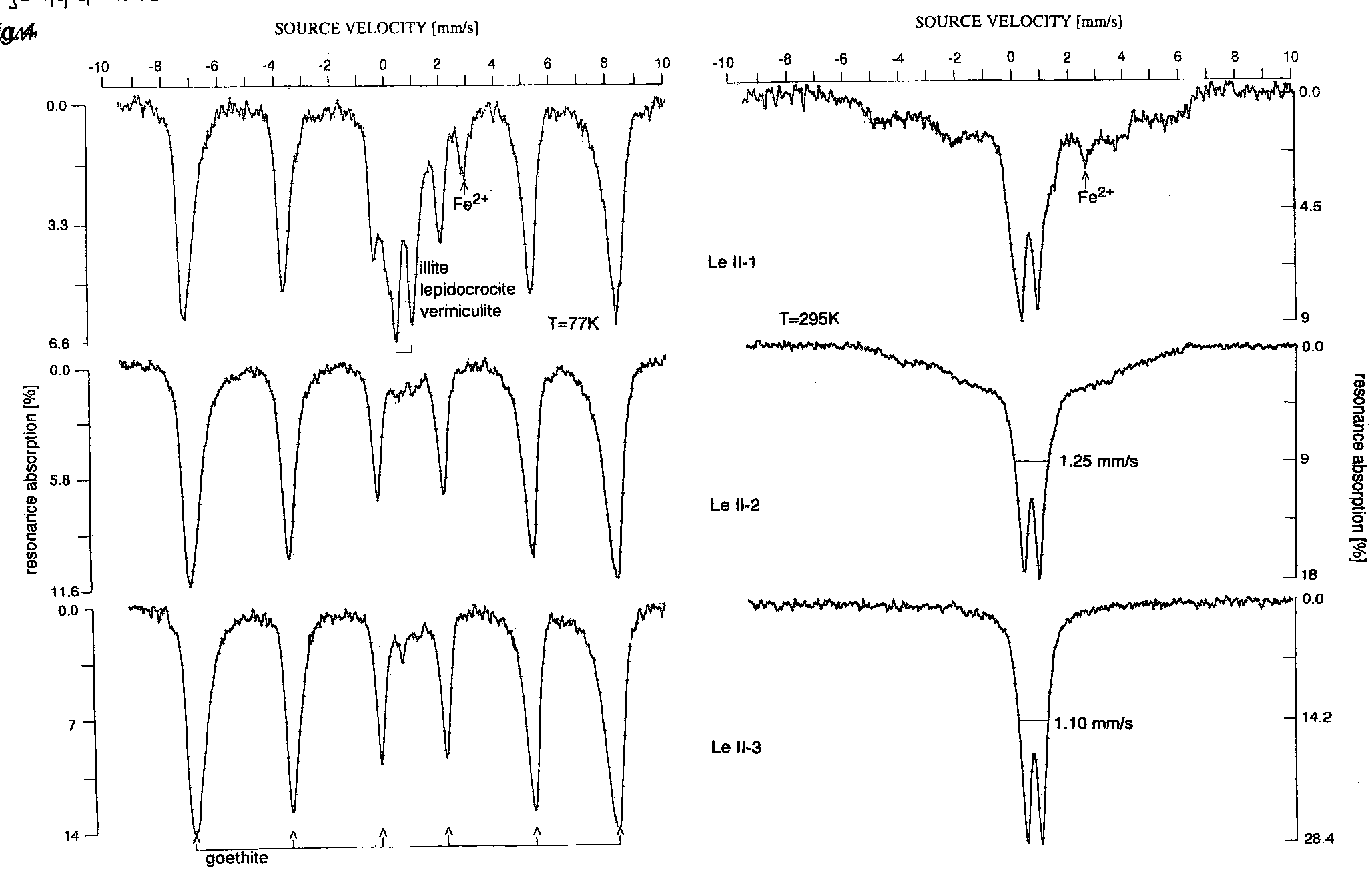
MS[295K] and MS[77K] of samples Le II are
shown in Figure 4. The spectra have much in com-mon with those of Ko II, particularly with respect tothe magnetic properties of goethite. The hyperfinefield B’[77K] is highest in Le II-1 and lowest in Le II-3 (Table 1). The non-magnetic part in MS[77K] ofLe II-1 is different, however, as it consists of threesubspectra, a single Fe2+ doublet and two Fe3+ dou-blets. One of the latter can be attributed to lepi-docrocite that orders magnetically just below 77 K.
Fig. 3. The magnetic hyperfine field, B’, and the hyperfine fraction,
This is verified by a measurement of MS at a temper-
h.f.f., in the Mössbauer spectra of Ko II-1, Ko II-2 and Ko II–3 as
ature of 4.2 K. Instead of a single sextet of goethite, a
a function of temperature. The full curve refers to the hyperfine
second one was found with a hyperfine field charac-
field in a goethite crystal; the dashed curve refers to B’ in synthetic
teristic for lepidocrocite at 4.2 K; XRD confirmed
goethite nanocrystals with a mean width of 9 nm. Dot-dash lines
this, and illite and vermiculite were identified in addi-
through the experimental data points of the Ko samples are drawn
tion (Table 2), contributing to the other two doublets
Fig. 4. Mössbauer spectra of samples Le II-1(core), Le II-2 and Le II–3 (both mantle) at 77 and 295 K. A central doublet dominates
MS[295K] that has practically disappeared in MS[77K], where six-line spectra attributed to goethite are most pronounced. The positions of
the lines that correspond to the hyperfine field, B’, are indicated. The central contribution to MS[77K] of Le II-1 can be assigned to lepi-
docrocite, illite and vermiculite. The FWHM of the central doublet in MS[295K] is given; note the increase of its line width in the presence
Geologie en Mijnbouw / Netherlands Journal of Geosciences 79(1) 2000
Table 3. Major oxides and a number of trace elements in samples from various rattlestones and loam from Koningsbosch, as obtained by
in MS[77K]. Temperature dependencies of both B’
and core of one of the central segments, Re-1 and Re-
and h.f.f. in samples Le II (Fig. 5) show the same
1a, and from consecutive shells of the siderite concre-
trend as found in Ko II. MS data on Le II suggest
tions Re-2 through 4. Goethite is the dominant iron
that the crystallite size of goethite is largest in the core
oxide in all samples. Siderite was identified in Re-2
and smallest in the exterior part of the mantle.
only and hematite was present in Re-1 and Re-1a.
The results of Ko II and Le II are representative for
The goethite crystallinity becomes worse in the shells
the other rattlestones. Goethite is the predominant
outwards, in the same way as in the mantle of rattle-
iron oxide in all of them, in both core and mantle. The
stones. Hematite in association with goethite has been
goethite-crystallite size is in the nanometer range,
found in several siderite concretions (Meyer, 1979;
which follows from its superparamagnetic behaviour
Senkayi et al., 1986; unpublished results from the au-
at 295 K and the dramatic lowering of the magnetic
thor); hematite has, however, not been identified in
ordering temperature, and is also consistent with what
is known from a comparison among common soilminerals (Schwertmann, 1988a). In general, the
goethite-crystallite size is smallest and tends to bemost heterogeneous in the exterior part of the mantle.
In order to verify the Mössbauer results, X-ray dif-
MS were obtained from samples of both surface
fractograms were obtained from several samples.
Geologie en Mijnbouw / Netherlands Journal of Geosciences 79(1) 2000
Fig. 5. The magnetic hyperfine field, B’ ,
and the hyperfine fraction, h.f.f., in the
ture. The full curve refers to the hyper-
fine field in a goethite crystal. Dot-dash
Table 1 lists peak positions and line widths of
in several samples from rattlestones found at sites
goethite reflections, and Table 2 lists the mineralogy.
north of the Meuse river (Table 2). Lepidocrocite
The peak positions are similar to those of pure
was mostly encountered in the mantle, except in Le
goethite, which makes isomorphic substitution of Fe
II-1. The line width of the (020) reflection is reason-
by Al unlikely. The (020), (110) and (111) lines and
ably narrow and hence lepidocrocite is more crys-
the average of the (130), (021), (121) and (140) lines
talline than goethite in these samples. Moreover, this
of goethite were chosen to investigate line broaden-
iron oxide is rarely evenly distributed over the whole
ing. Although the first three lines in the core samples
soil matrix (Schwertmann, 1988b). This might ex-
were subject to interference with strong quartz lines,
plain why lepidocrocite was found in some laminae
the other four lines are not. A systematic increase in
but is absent in adjacent ones. Lepidocrocite is not
line broadening is observed in the samples Ko II and
found in rattlestones from sites east of the Meuse, al-
Le II from the core outwards, as shown in Table 1,
though Riezebos et al. (1978) suggested that this iron
indicating a decrease in crystallinity (Schwertmann,
oxide is a constituent in middle terraces in south
1988a), consistent with MS data. XRD data on line
broadening in goethite in the mantles of Ko III and
XRD results on clay mineral identification are given
La show a similar trend. The goethite content in the
in Table 2, where they are also compared with MS re-
core of these rattlestones was too low to determine
sults. The overall correspondence is good. Besides
the former. The weight percentage of goethite in each
goethite, illite is found in most samples, not surpris-
sample was derived from the absolute Fe content,
ingly, as it is the main clay mineral in Dutch soils
determined by INAA, in combination with the
(Edelman & De Bruin, 1986). Vermiculite, identified
goethite fraction measured in MS[77K]. In addition,
by XRD in some core samples, can be associated
the temperature of onset of magnetic ordering in
with a relatively high Fe2+ signal in MS. Table 2 also
goethite, T , is presented in Table 1. The lower this
lists the XRD results on the siderite concretion. With
temperature, the poorer the goethite crystallinity
respect to the iron compounds they are consistent
(Murad & Bowen, 1987) consistent with the stronger
with MS. All Re samples contain dolomite, which is
absent in the rattlestone samples. Clay minerals were
In both MS and XRD, lepidocrocite was identified
encountered only in the shells of the concretion.
Geologie en Mijnbouw / Netherlands Journal of Geosciences 79(1) 2000
trace elements show a similar trend: the amount ofRb and Cs, for instance, is positively correlated with
Elemental composition of the samples has been ob-
illite, which is consistent with previously reported re-
tained by INAA. The Fe content in the core varies
sults (Moura & Kroonenberg, 1990).
from 18% by weight in S II to 1.7% in De I; the latter
Heavy metals can, as a rule, be associated with Fe
has hardly any Fe accumulation at all. In contrast, the
hydroxides – supplied by groundwater – that remain
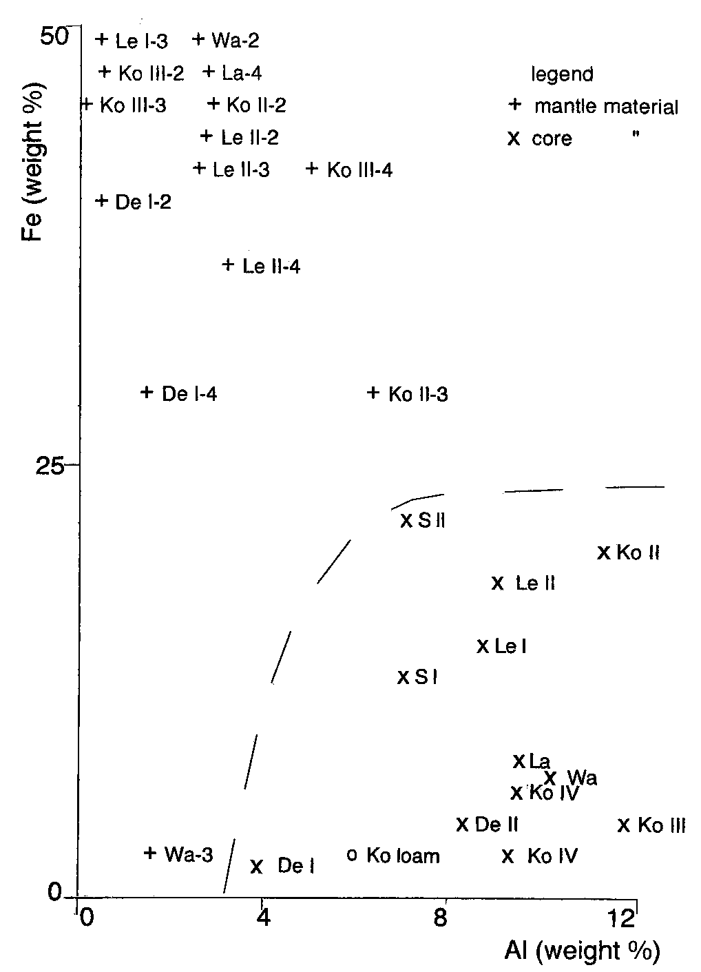
Fe content in the core segment of the siderite concre-tion is 52% by weight. The mantle of rattlestones hasa high iron accumulation (up to 50% by weight) thatvaries by less than a factor of 2 among the varioussamples (except for Wa-3). The Fe content relates tothat of Al as shown in Figure 6; in general, a high con-tent in the former corresponds to a low content in thelatter and vice versa. The amount of Fe gradually de-creases in each rattlestone in consecutive layers of themantle outwards, while the content of various otherelements just increases. A histogram (Fig. 7) illus-trates how the total Fe content in core and mantle offour different rattlestones is distributed amonggoethite, lepidocrocite and clay minerals, as deter-mined in MS. Goethite is the predominant iron ox-ide; lepidocrocite has been identified only in the rat-tlestones from north of the Meuse river (De I and LeII). The composition in terms of major oxides (inweight percentage) and a few trace elements (in ppm)is presented in Table 3.
The amount of silicon in the core is about twice
that in the mantle, which is understandable as themuch higher Fe accumulation in the mantle lowersthe relative Si content in the latter. A quantitative esti-mate of illite has been made, assuming that K is fullyassociated with this clay mineral by using the chemi-cal formula, KAl Si O H . The illite content in the
Fig. 7. A plot of the total Fe versus Al content, obtained by INAA,
core in general exceeds that in the mantle by a sub-
in core and mantle of the rattlestones (indicated by their codes).
stantial factor, and it tends to increase gradually out-
Below the dotted line are the Fe and Al content of the core sam-
wards in the latter. This could be a reason why several
ples. Clayey loam from the Koningsbosch site is indicated by (o).
Geologie en Mijnbouw / Netherlands Journal of Geosciences 79(1) 2000
incorporated in the iron oxide after sedimentation.
in the Netherlands (Huisman et al., 1999). It appears
The element Ba is also positively correlated with illite
that the pattern in the mantle is similar to that in
and the ratio Ba(ppm)/K O is similar to the value for
groundwater. The relative depletion of LREE in the
subsurface sediments in the southern Netherlands
mantle is attributed to the fact that they form less eas-
(Huisman et al., 1997). In the samples Ko II-2, Ko
ily carbonate complexes than HREE, due to their
II–3, Ko III-2 and Ko III–3, however, Ba is very
larger ionic radii. It is known that bicarbonate ion
strongly accumulated (Table 3). Such irregular accu-
pairs can play an important role for the solution
mulations of Ba without correlation with illite or Fe
chemistry of HREE, which explains a significant rela-
may indicate that it is present as a separate mineral
tive fractionation and an enrichment in the latter
phase or that it is built into the Fe-oxide structure in
(Michard et al., 1987). The pattern in the core, on the
varying ratios to iron (Huisman, 1998). Samples with
other hand, is more shale-like without significant frac-
an excessively high Ba content show a high Mn con-
tionation, which is more common for river water
tent, too. It follows also from Table 3 that the distribu-
(McLennan, 1989). In summary, the mantle has been
tion of Th and U in core and mantle is different; the
formed from elements that are mainly supplied by
mean ratio Th/U amounts to 4 in the core and is
groundwater, whereas those in the core originate pri-
about 1 in the mantle. These elements tend to frac-
marily from the river water. In this connection it is
tionate because of oxidation of U to soluble ions, and
noted that loam from Koningsbosch also has a shale-
because of selective adsorption of Th in clays and its
retention in heavy resistant minerals (Wedepohl,
Variations in the content of heavy metals within the
mantle and between various rattlestones are probably
The mean content of the rare earth elements in the
caused by differences in groundwater composition
mantle is systematically higher than in the core, ex-
during the formation of these stones. Such diverse-
cept for La, Ce and Nd, which elements show an op-
ness could be the result of differences in aquifer mate-
posite behaviour. The shale-normalised REE patterns
rial, and by changes in pH and Eh of the ground-
for both core and mantle are shown in Figure 8 and
compared with that reported for shallow groundwater
The Munsell hue of the samples, using the Munsellsoil color charts (1954) is also indicated in Table 2. The color of the iron oxide may vary from sample tosample and often has sufficient consistency to be use-ful for the identification of these oxides in soils andsediments (Schwertmann, 1988a). The mineral-spe-cific Munsell hues of iron oxides in soils are in the fol-lowing ranges: hematite 5R-2.5YR, lepidocrocite 5YR-7.5YR, and goethite 7.5YR-2.5Y, sometimes ex-tending to 5YR.
Finely dispersed soil goethite and lepidocrocite
usually become increasingly dark with poorer crys-tallinity; goethite crystals smaller than 50 nm, for in-stance, are brownish (Cornell & Schwertmann,1996). The first number behind the color hue, calledthe value, is a notation for darkness varying between 0(absolute black) and 8 (absolute white). Core sampleshave a higher value than those of the mantle, which isqualitatively consistent with the better crystallinity ofgoethite in the former. Discussion
Fig. 8. REE patterns in core and mantle of rattlestones obtained by
Goethite forms in all soil types and in early stages of
INAA are compared with the patterns found in shallow groundwa-
weathering it is the most commonly found iron oxide.
ter and in a sandpit. The concentrations are normalised using
Dissolved ferrous iron, which is usually present in re-
Geologie en Mijnbouw / Netherlands Journal of Geosciences 79(1) 2000
duced sediments, oxidizes to Fe(III) when it encoun-
pects: (1) the iron content in the central segments of
ters oxygen and converts to virtually insoluble Fe(III)
the concretion is very high and clay minerals are ab-
oxyhydrates (Cornell & Schwertmann, 1996). In al-
sent in contrast to what is found in the core of rattle-
ternating reducing and oxidizing conditions, a clay
stones, (2) hematite is found in the central segments
layer in a sandy matrix will accumulate these hydrates
and absent in the rattlestone samples, (3) the latter
through the following process. When the sediment
may contain a strong and sometimes irregular accu-
layer is reduced, Fe(II) is distributed evenly in the
mulation of heavy metals such as Ba, which is unlikely
aqueous phase. When this sediment dries out, iron
in siderite as its structure cannot accommodate any
will partially precipitate, being exposed to oxygen,
accumulation of heavy metals (Huisman, 1998), and
while the concentration in the remaining solution in-
(4) the specific density of rattlestones can, due to a
creases. Eventually the finer layers will still contain
large cavity, be much smaller than the densities re-
water and its dissolved iron. Upon complete dryout,
ported by Van der Burg (1969), as shown in Figure 1.
this iron precipitates at the contact of the fine layer
Septarian cracks have indeed been found in the alter-
(which is still water-saturated) and its aerated coarse
ated siderite concretion, which result in a relatively
surroundings. This is a repetitive process. In addition
small cavity only. These differences make it unlikely
to accumulate iron, the clay layer may fracture upon
that rattlestones are the result of oxidation of siderite.
drying out, whereby each of the fragments will be-
It examplifies what Fitzpatrick (1988) has described
come a nucleus of Fe accumulation. With time, the
as iron compounds being indicators of pedogenic
accumulated iron may form a rigid oxide coating
around the clay core, and become a concretion. When
Rattlestones are composed of clay minerals, mainly
the concretion dries out further, the clay core will
illite and vermiculite, and poorly crystalline goethite.
shrink and detach from the mantle, thus forming a
The main non-clay mineral is quartz. This mixture
cavity in the stone. Because the inner part of the rat-
closely corresponds with what was called an assem-
tlestone is not subject to periodical reduction and
blage of young, moderately weathered soils in temper-
precipitation any more, it can recrystallize to larger
ate regions by Van Breemen & Buurman (1998). The
size, while this is not the case for the active outer part
official definition of a rattlestone given in the intro-
(Van Breemen & Buurman, 1998). These processes
duction (Jackson, 1997) will be more in accordance
are governed throughout the year by groundwater so
with the actual results when it is recognized that the
that absolute accumulation of iron, conveyed from
core material does not consist of loose sand only.
surrounding areas, may occur. As a result of fluctuat-
The elemental analysis of several samples of a rat-
ing groundwater levels, yellowish brown goethite con-
tlestone gives additional geochemical information on
centrates form laminae in the mantle of the stone
the subsurface soil and sediments of the sampling
with an Fe(III)-oxide content that is high relative to
area. For example, the higher accumulation of various
the surroundings. These oxides may cement the min-
heavy metals in rattlestones from the older flood-
eral grains giving rise to the hardness of the concre-
plains east of the Meuse river compared to that in
stones from north of the river indicates a richer min-
The size of the cavity will depend on the amount
eralogy in the former. On the other hand, the absence
and mineralogy of the clay, and the original water
of lepidocrocite in rattlestones from Koningsbosch
content. Since illite is the dominant clay mineral, its
might imply that this iron oxide has been transformed
content in the core is positively correlated with the
into goethite in the course of time though this process
size of the cavity in the rattlestones as can be seen by
is very slow under pedogenic conditions (Schwert-
mann & Taylor, 1972). As lepidocrocite occurs pre-
The former process of genesis of rattlestones is en-
dominantly in younger postglacial soils following
tirely different from the suggestion by Van der Burg
Schwertmann & Taylor (1971), the reason for its ab-
(1969) that they have been formed as a result of the
sence is because these rattlestones are from the much
oxidation of siderite concretions that had been de-
earlier Sterksel Formation (Middle Pleistocene). Lepi-
posited contemporaneously with the sediments in the
docrocite was not found in the siderite concretion.
beds where they are found today. As siderite is particu-larly vulnerable to weathering under atmospheric
Conclusions
conditions, it will oxidize preferentially to goethite(Pettijohn, 1975). Although MS and XRD results
The chemical and mineralogical composition of rat-
with respect to goethite crystallinity in the siderite
tlestones found near the main Dutch rivers has been
concretion are qualitatively similar to those obtained
extensively studied by Mössbauer spectroscopy,
with rattlestones, both systems differ in several as-
INAA and XRD. Rattlestones are iron concretions
Geologie en Mijnbouw / Netherlands Journal of Geosciences 79(1) 2000
formed in an environment of lateral accumulation of
of the Mössbauer group in the Department of Radia-
Fe under the influence of periodical oxidation,
tion Physics (IRI) and to Ing. M.P. van Steenvoorden
around a fine core of ferruginous sediments, mainly
for giving me the opportunity to carry out the Möss-
clay and sand. The core has shrunk and detached it-
self from the mantle around it. The cavity formed canhave a considerable size, of up to 50% by volume. References
57Fe Mössbauer spectroscopy was applied to identify
Adams, F.D., 1938. The birth and development of the geological
the iron oxides, among which goethite is predomi-
sciences. Ballière, Tindall and Cox (London): 506 pp.
nant. The goethite crystallinity was investigated by
Bromehead, C.N., 1947. Aetites or the eagle-stone. Antiquity 21:
making use of its magnetic properties and showed to
be poorest in material from the outer edge of the rat-
Coey, J.M., 1988. Magnetic properties of iron in soil iron oxides
and clay minerals. In: Stucki, J.W., Goodman, B.A. & Schwert-
tlestone. The latter is confirmed by broadening of the
mann, U. (eds.): Iron in soils and clay minerals. D. Riedel (Dor-
various X-ray reflections. In addition, illite and vermi-
culite were identified by XRD; they were found main-
Cornell, R.M. & Schwertmann, U., 1996. The iron oxides: struc-
ture, properties, reactions, occurrence and uses. Verlag Chemie
The elemental composition was determined by
Edelman, Th. & De Bruin, M., 1986. Background values of 32 ele-
INAA. The iron content in the mantle is about 50%
ments in Dutch topsoil, determined with non-destructive neu-
by weight and gradually decreases outwards, while
tron activation analysis. In: Assink, J.W. & Van den Brink, W.J.
the core contains 2-15% Fe by weight. Differences
(eds.): Contaminated soil. Martinus Nijhoff Publishers (Dor-
between rattlestones from a Middle Pleistocene site
east of the Meuse river and those from Late Pleis-
Fitzpatrick, R.W., 1988. Iron compounds as indicators of pedo-
genic processes: examples from the southern hemisphere. In:
tocene sites north of the river are the absence of lepi-
Stucki, J.W., Goodman, B.A. & Schwertmann, U. (eds.): Iron in
docrocite and a richer mineralogy in the former. As a
soils and clay minerals. D. Riedel (Dordrecht): 351-395.
result of fluctuating groundwater levels, iron and oth-
Huisman, D.J., 1998. Geochemical characterization of subsurface
er (trace) elements were supplied and could lead to
sediments in the Netherlands. Thesis Agricultural University Wa-
concentrates, of iron hydroxides in particular, in the
Huisman, D.J., Vermeulen, F.J.M., Baker, J., Veldkamp, A., Kroo-
most porous parts of the clayey sediment. The de-
nenberg, S.B. & Klaver, G.Th, 1997. A geological interpretation
tailed mechanism describing the formation of rattle-
of heavy metal concentrations in soils and sediments in the
stones is consistent with experimental results. Its gen-
southern Netherlands. Journal of Geochemical Exploration 59:
esis is from inside outwards and is difficult to recon-
cile with the concept of oxidation of a siderite concre-
Huisman, D.J., Van Os, B.J.H., Klaver, G.Th. & Van Loef, J.J.,
1999. Redistribution of REE in aquifers in the Netherlands. In:
Ármannsson, H. (ed.): Geochemistry of earth’s surface. A.A. Acknowledgements
Jackson, J.A., 1997. Glossary of geology (4th ed.). American Geo-
logical Institute (Alexandria): 769 pp.
I like to express my high appreciation for the fruitful
Kuzmann, E., Nagy, S., Vértes, A., Weiszburg, T.G. & Garg, V.K.,
1998. Geological and mineralogical applications of Mössbauer
discussion with and valuable support by Dr. P. Buur-
spectroscopy. In: Vértes, A., Kuzmann, E., Nagy, S., Weiszburg,
man and J.D.J. Van Doesburg for the XRD measure-
T.G. & Garg, V.K. (eds.): Nuclear methods in mineralogy and
ments in the Laboratory of Soil Science and Geology
geology. Plenum Press (New York): 285-376.
at the Wageningen University. Discussions with Dr.
McLennan, S.M., 1989. Rare earth elements in sedimentary rocks:
H. Huisman (NITG-TNO) on REE have been very
influence of provenance and sedimentary processes. In: Lipin,
B.R. & McKay, G.A. (eds.): Geochemistry and mineralogy of
useful. I have profited from the generosity of readers
rare earth elements. Reviews in Mineralogy 21: 169-200.
of the Dutch journal of the Nederlandse Geologische
Meyer, B., 1979. Die Entcarbonatierungsrotung als Bodengeneti-
Vereniging, Grondboor en Hamer, for providing me
scher Teilprozess. Mitteilungen der deutschen Bodenkundlichen
with a number of rattlestones. I am grateful to the late
Ir. C. Maugenest of the Mineralogical and Geological
Michard, A., Beaucaire, C. & Michard, G., 1987. Uranium and
rare earth elements in CO -rich waters from Vals-les Bains
Museum, Department of Applied Geoscience of
(France). Geochimica et Cosmochimica Acta 51: 901-909.
Delft University of Technology, and to Dr. T. G. Nij-
Moura, M.L. & Kroonenberg, S.B., 1990. Geochemistry of Qua-
land, who helped me in an early stage of the research.
ternary fluvial and eolian sediments in the southeastern Nether-
I appreciate the vivid interest of Drs. B.W. Zuurdeeg
lands. Geologie en Mijnbouw 69: 359-373.
from Geochem Research (Utrecht) in this study. I
Munsell Soil Color Charts, 1954. Munsell Company Inc. (Balti-
thank Mrs. Drs. Th.G. van Meerten (Radiochemistry
Murad, E. & Bowen, L.H., 1987. Magnetic ordering in Al-rich
Department of IRI at Delft) for carrying out INAA. I
goethites: influence of crystallinity. American Mineralogist 72:
am very thankful to Dr. Ir. A.M.Van der Kraan, head
Geologie en Mijnbouw / Netherlands Journal of Geosciences 79(1) 2000
Murad, E., 1988. Magnetic properties of iron in soil iron oxides
tion of soil lepidocrocite to goethite. In: Pseudogley and gley.
and clay minerals. In: Stucki, J.W., Goodman, B.A. & Schwert-
Transactions of Committees V and VI of the International Soci-
mann, U. (eds.): Iron in solids and clay minerals. D. Riedel (Dor-
ety of Soil Science (Stuttgart-Hohenheim): 45-54.
Schwertmann, U. & Taylor, R.M., 1972. The transformation of lep-
Murad, E., 1996. Magnetic properties of microcrystalline iron (III)
idocrocite to goethite. Clays and Clay Minerals 20: 151-158.
and related materials as reflected in their Mössbauer spectra.
Senkayi, A.L., Dixon, J.B. & Hossner, L.R., 1986. Todorokite,
Physics and Chemistry of Minerals 23: 248-262.
goethite and hematite: alteration products of siderite in East
Parry, S.J., 1991. Activation spectrometry in chemical analysis.
Texas lignite overburden. Soil Science 142: 36-42.
J.Wiley & Sons (NewYork) 243 pp.
Van Breemen, N. & Buurman, P., 1998. Soil formation. Kluwer
Pettijohn, F.J., 1975. Sedimentary rocks. Harper & Row (New
Academic Publishers (Dordrecht): 377 pp.
Van der Burg, W.J., 1969. The formation of rattle stones. Palaeo-
Riezebos, P.A., Bisdom, E.B.A. & Boersma, O., 1978. Composite
geography, Palaeoclimatology, Palaeoecology 6: 105-124.
grains in Maas sediments: a survey and a discussion of their
Van der Burg, W.J., 1971. The climatological factors. Palaeogeogra-
opaque components. Geologie en Mijnbouw 57: 417-431.
phy, Palaeoclimatology, Palaeoecology 7: 297-308.
Schwertmann, U., 1988a. Some properties of soil and synthetic
Van der Kraan, A.M., 1972. Mössbauer effect studies of superpara-
iron oxides. In: Stucki, J.W., Goodman, B.A. & Schwertmann, U.
magnetic α-FeOOH and α-Fe O . Thesis Delft University of
(eds.): Iron in solids and clay minerals. D. Riedel (Dordrecht):
Van der Kraan, A.M. & Van Loef, J.J., 1966 Superparamagnetism
Schwertmann, U., 1988b. Occurrence and formation of iron oxides
and submicroscopic α-FeOOH particles observed by the Möss-
in various pedoenvironments. In: Stucki, J.W., Goodman, B.A. &
bauer effect. Physics Letters 20: 614-616.
Schwertmann, U. (eds.): Iron in solids and clay minerals. D.
Wedepohl, K.H. (ed.), 1978. Handbook of geochemistry. Springer
Schwertmann, U. & Taylor, R.M., 1971. The in vitro transforma-
Geologie en Mijnbouw / Netherlands Journal of Geosciences 79(1) 2000
 source velocity in Mössbauer spectroscopy. TheMössbauer spectrum at 295 K (coded as MS[295K])of paramagnetic lepidocrocite consists of two lines ofequal intensity, the quadrupole splitting. The doubletis much larger in MS of siderite and readily distin-guished from that of lepidocrocite. Speciation in asample containing a few percent of Fe by weight isfeasible. Poorly crystalline iron oxides can still beidentified, in particular at lower temperatures. Spec-tral intensities of iron-containing species give infor-mation on their relative content in a sample. Furtherdetails on Mössbauer spectroscopy can be found inKuzmann (1988).
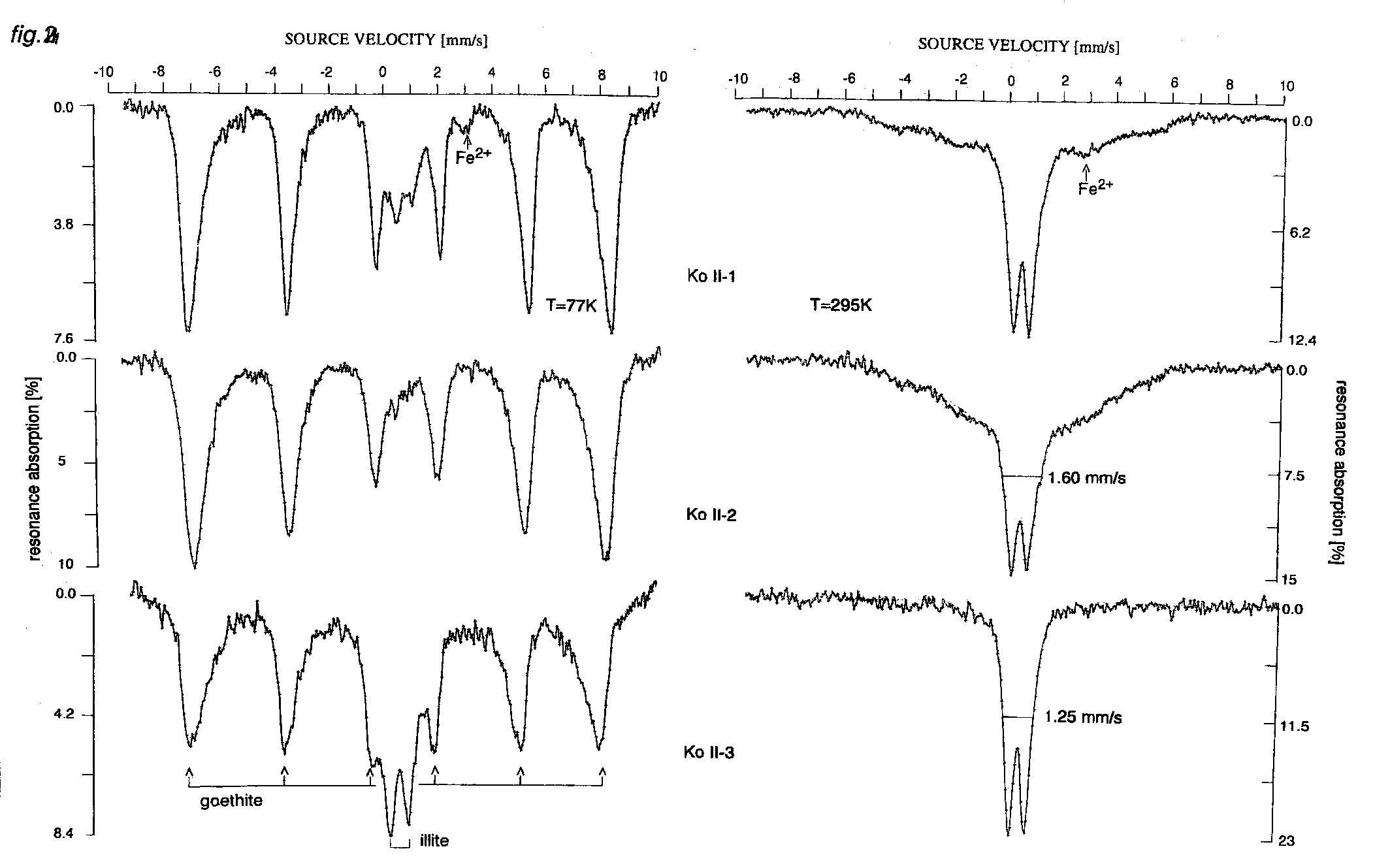 Fig. 2. Mössbauer spectra of samples Ko II-1 (core), Ko II-2 and Ko II–3 (both mantle) at 77 and 295 K. A central doublet dominates
MS[295K] that has practically disappeared in MS[77K], where six-line spectra attributed to goethite are most pronounced. The positions of
the lines that correspond to the hyperfine field, B’, are indicated. A central doublet in MS[77K] of Ko II-1 and Ko II–3 can be ascribed to il-
lite, including the very small Fe2+ signal . The illite also contributes to the central doublet in MS[295K] but coincides with that of superpara-
Table 1. Characteristics of the various samples as determined by XRD and Mössbauer spectra. The line width has not been corrected for in-
strumental resolution of 0.1°. The goethite content in the core of Ko III and La was too low to measure the reflections.
Fig. 2. Mössbauer spectra of samples Ko II-1 (core), Ko II-2 and Ko II–3 (both mantle) at 77 and 295 K. A central doublet dominates
MS[295K] that has practically disappeared in MS[77K], where six-line spectra attributed to goethite are most pronounced. The positions of
the lines that correspond to the hyperfine field, B’, are indicated. A central doublet in MS[77K] of Ko II-1 and Ko II–3 can be ascribed to il-
lite, including the very small Fe2+ signal . The illite also contributes to the central doublet in MS[295K] but coincides with that of superpara-
Table 1. Characteristics of the various samples as determined by XRD and Mössbauer spectra. The line width has not been corrected for in-
strumental resolution of 0.1°. The goethite content in the core of Ko III and La was too low to measure the reflections.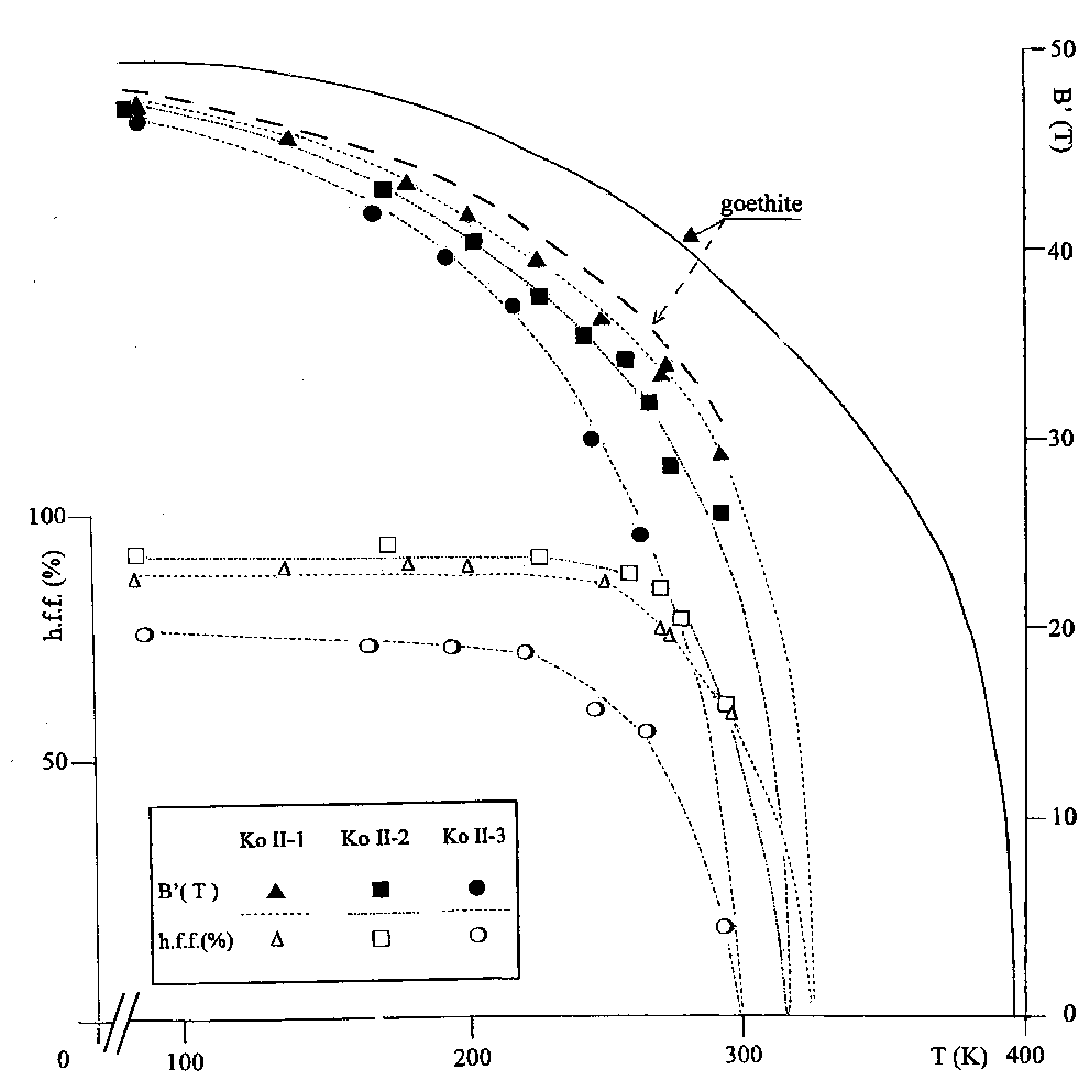
 MS. Figure 3 shows that Ko II-3 not only has thelowest temperature of onset of magnetic ordering ingoethite, but is also the least homogeneous with re-spect to goethite-crystallite size distribution, as thetemperature range in which magnetically ordered andsuperparamagnetic phases of goethite coexist iswidest. This corroborates the strongly broadened hy-perfine lines in MS[77K] of Ko II-3.
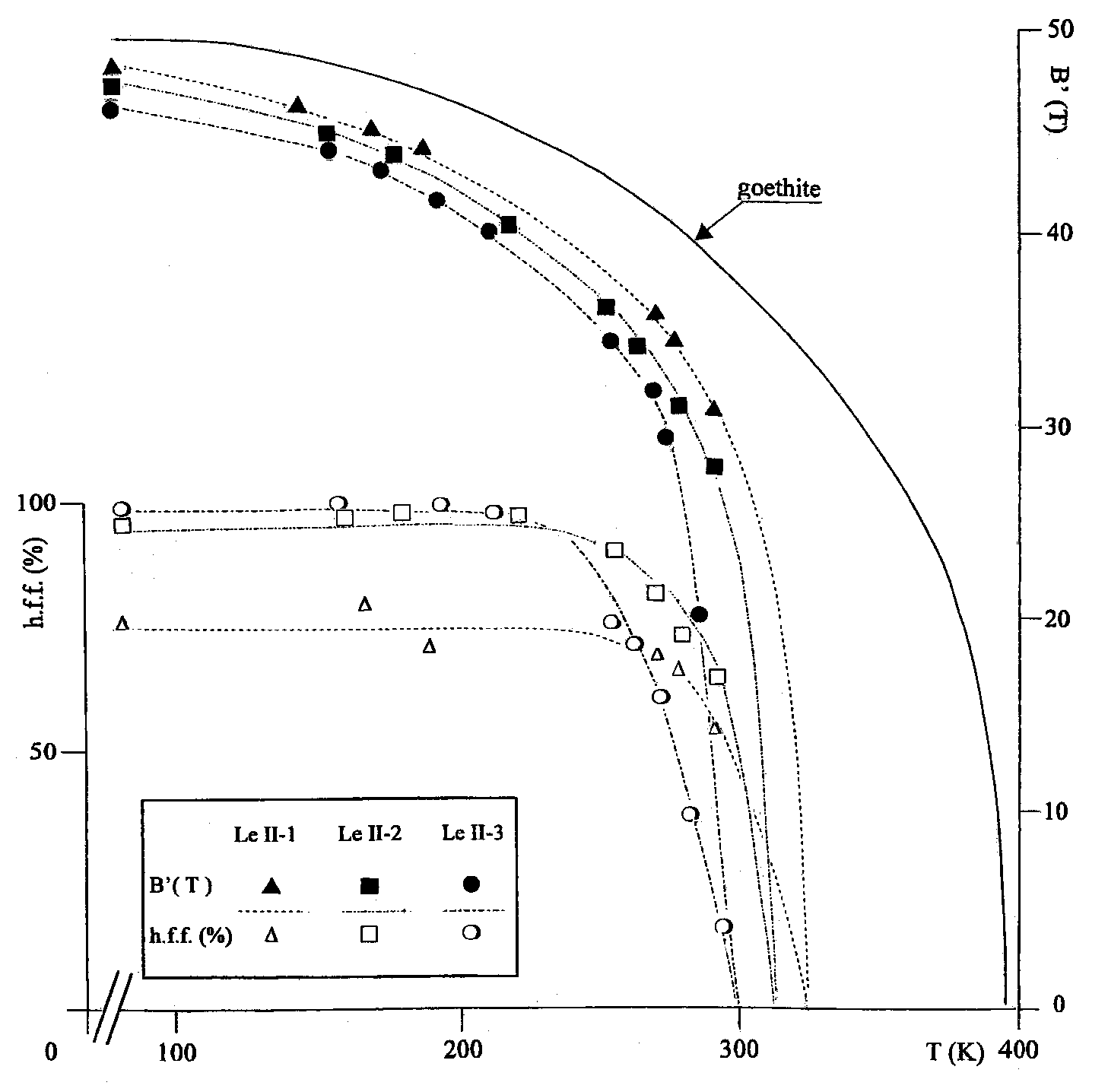 Fig. 5. The magnetic hyperfine field, B’ ,
and the hyperfine fraction, h.f.f., in the
ture. The full curve refers to the hyper-
fine field in a goethite crystal. Dot-dash
Table 1 lists peak positions and line widths of
in several samples from rattlestones found at sites
goethite reflections, and Table 2 lists the mineralogy.
Fig. 5. The magnetic hyperfine field, B’ ,
and the hyperfine fraction, h.f.f., in the
ture. The full curve refers to the hyper-
fine field in a goethite crystal. Dot-dash
Table 1 lists peak positions and line widths of
in several samples from rattlestones found at sites
goethite reflections, and Table 2 lists the mineralogy.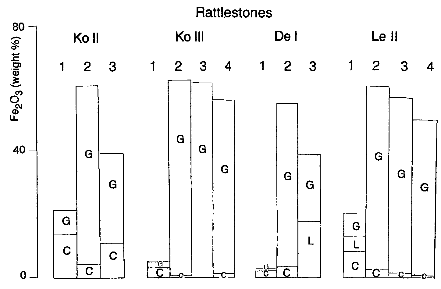
 trace elements show a similar trend: the amount ofRb and Cs, for instance, is positively correlated with
Elemental composition of the samples has been ob-
illite, which is consistent with previously reported re-
tained by INAA. The Fe content in the core varies
sults (Moura & Kroonenberg, 1990).
trace elements show a similar trend: the amount ofRb and Cs, for instance, is positively correlated with
Elemental composition of the samples has been ob-
illite, which is consistent with previously reported re-
tained by INAA. The Fe content in the core varies
sults (Moura & Kroonenberg, 1990).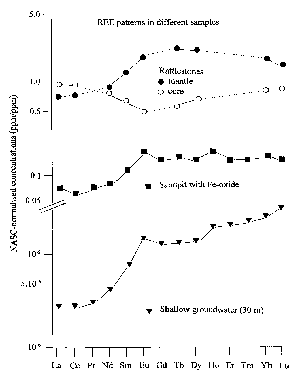 incorporated in the iron oxide after sedimentation.