Ec113 120.123

Exp Clin Endocrinol Diabetes 109 (2001) 120 ± 123 Fatigue and hyponatremia in a 75-year-old woman: unusual presentation of hypophysitisJ. Klein, W. Kern, H. L. FehmDepartment of Internal Medicine I, Medical University of Lübeck, Germany Key words: Hypophysitis, hyponatremia, hypopituitarism secondary hypothyroidism. Hormonal substitution with hydro- Summary: A 75-year-old woman presented with general fatigue cortisone and levothyroxine resulted in rapid improvement of all progressing to somnolence. Laboratory tests showed marked symptoms and signs. Without additional treatment shrinkage of the hyponatremia, TSH in the normal range, but low levels of free T3 pituitary gland could be documented. Our report extends the and free T4. Evaluation of pituitary hormones and magnetic known clinical and pathological spectrum of hypophysitis and resonance imaging of the pituitary unmasked findings character- illustrates the need to include this uncommon entity in the istic for hypophysitis with secondary adrenal insufficiency and differential diagnosis of hyponatremia even in elderly patients.
regular medication. Except for the treatment of Hypophysitis is a rare inflammatory disorder, first tuberculosis forty years ago, the past history was described by Goudie in 1962, which may lead to destruction of the anterior pituitary and usually On examination, the patient appeared somnolent, affects young women in late pregnancy or postpartum but fully oriented. She was 1.63 m tall and weighed (Beressi et al., 1999; Goudie and Pinkerton, 1962).
56 kg. She was afebrile, her blood pressure was 180/ Histological examination classically reveals infiltra- 90 mmHg, the heart rate was 80 beats/min and tion of the pituitary tissue by lymphocytes and regular. Her face was pale and appeared slightly macrophages (Beressi et al., 1999), a finding that has puffy, skin turgor was normal, there were no signs of led to the coining of the term lymphocytic hypo- oedema and no pathological findings on examination physitis. Typical features of the clinical presentation of the lungs, heart and abdomen. All peripheral include headaches, hypopituitarism with predomi- pulses were palpable. Neurological examination nant symptoms and signs of adrenal insufficiency, and revealed no abnormality of the cranial nerves, visual field loss (Beressi et al., 1999; Cosman et al., particularly no visual field deficits. Apart from 1989; Jenkins et al., 1995; Powrie et al., 1995). Here, symmetrical distal hypaesthesia in both feet and we describe the unusual case of a 75-year-old woman diminished ankle reflexes, there were no signs of with general fatigue and hyponatremia as only prominent features that turned out to be caused by The results of routine hematological, blood chem- secondary adrenal insufficiency and hypothyroidism ical and enzyme tests were normal with the exception of a marked hyponatremia and elevated values for C- reactive protein, fibrinogen and CK (Table 1). TSH was in the normal range, but free T3 and T4 were decreased. Furthermore, anti-TPO antibody levels The electrocardiogram, a chest radiograph and an A 75-year-old woman presented with complaints of abdominal ultrasound showed no significant abnor- increasing fatigue and general loss of energy that had malities. Magnetic resonance imaging of the brain progressively been developing over the last five days.
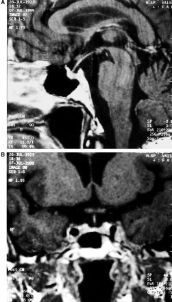
revealed a slightly enlarged pituitary that partially Furthermore, she had noticed a puffy face and felt extended above the sella. Gadolinium enhancement nauseous. She did not report weight loss, night sweats, showed homogeneous uptake in the entire pituitary or fevers, and the remaining functional enquiry was with a prominent rim along the sellar diaphragm unremarkable. She did not smoke and was on no J. Klein et al., Hypophysitis in a 75-year-old woman Table 1 Pathological blood chemical values at presentation Corticotropin-Releasing Hormone stimulation. Sim- ilarly, Thyrotropin-Releasing Hormone stimulation did not elicit an adequate response of TSH. Finally, secondary hypogonadism was detected as evidenced by low FSH and LH baseline levels and the absence of a significant rise of these hormones after Gona- dotropin-Releasing Hormone stimulation.
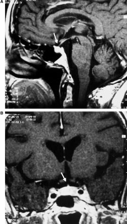
Treatment with 200 mg hydrocortisone per day and subsequently 50 mg levothyroxine resulted in rapid improvement of all symptoms. Sodium values re- turned to normal within the following four days with free water restriction only. Furthermore, the levels of plasma free T3 and T4 as well as basal cortisol also normalized. Magnetic resonance imaging of the pituitary three months later showed a rather small but otherwise normal appearing pituitary with only minimal enhancement of the sellar diaphragm (see Fig. 2). Seventeen months after the initial presenta- tion the patient is well. She is currently taking 100 mg levothyroxine and 30 mg hydrocortisone per day.
DiscussionIn this report, we describe a 75-year-old woman in whom fatigue and hyponatremia were the leading clinical and laboratory features at the first presenta- tion. The clinical course, evaluation of pituitary hormones, and imaging studies led to the diagnosis of hypophysitis causing secondary hypothyroidism and adrenal insufficiency. Not only the initial imaging characteristics ± demonstrating suprasellar extension of the pituitary and dural contrast enhancement described as pathognomonic ªdural tailº (Ahmadi et al., 1995; Honegger et al., 1997) ± but also the radiographic changes three months later ± charac- terized by an almost complete restitutio ad integrum ± corroborated the diagnosis. Furthermore, for hypo- Fig. 1 Contrast-enhanced computed tomography of the pituitary physitis, a preference for destruction of ACTH and gland in sagittal (A) and coronal (B) planes at first presentation. In TSH secreting cells has been reported (Beressi et al., both views there is enlargement of the pituitary gland which extends beyond the sellar diaphragm (arrow in A). The entire 1999; Jensen et al., 1986; Mayfield et al., 1980; pituitary takes up the contrast medium with accentuation of a small McDermott et al., 1988; Richtsmeier et al., 1980), a rim along the diaphragm (ªdural tailº, arrow in B) finding that is in exact concordance with our case.
Yet, a number of features of the case presented here Pituitary hormone tests and clinical course First, the age of onset of hypophysitis is very Results of pituitary stimulation tests are shown in unusual. Normally, the onset of this rare entity is seen Table 2. They revealed an inadequate response of most commonly in young women in late pregnancy ACTH and cortisol (from very low baseline levels) to or in the postpartum period with a mean age at Table 2 Results of pituitary function tests at presentation Plasma hormone levels were measured before (À60, À15), at the time (0), and after (+15, +30, +45, +60, +90) the application of an intravenous bolus injection of 60 mg CRH, 1.5 mg LHRH, and 12 mg TRH; range of normal values in parenthesis presentation of 31 years (Beressi et al., 1999; Powrie et al., 1995). To our knowledge, this is the first report of a woman 75 years of age presenting with hypophysitis. Interestingly, we also found elevated TPO-antibodies in the patient presented. This is in keeping with previous reports (Barbaro and Loni, 2000; Beressi et al., 1999; Goudie and Pinkerton, 1962; Nagai et al., 1997; Pestell et al., 1990; Sobrinho- Simoes et al., 1985) and lends support to the hypothesis of an autoimmune origin of hypophysitis.
Second, in contrast to the typical clinical presen- tation with headaches ± that often appear inappropri- ately severe for the degree of pituitary enlargement (Beressi et al., 1999; Honegger et al., 1997; Ikeda and Okudaira, 1987; Meichner et al., 1987) ± our patient only reported fatigue as her main complaint. This symptom as well as the laboratory finding of hyponatremia can be explained by a combination of secondary adrenal insufficiency and secondary hypothyroidism. In this context, elevation of CK indicates the presence of hypothyroidism-induced myopathy. The case of our patient illustrates that in elderly individuals, hypophysitis may present differ- ently. Especially in patients in this age group, many non-specific complaints are frequently encountered.
As highlighted by the findings in this case, serum TSH alone may not be sufficient in the laboratory work up of some patients since central hypothyroidism can Finally, treatment with physiological doses of hydrocortisone and levothyroxine substitution re- sulted in marked and sustained improvement of the patient with normalization of pituitary size. Current- ly, no uniform treatment recommendations for hypophysitis exist. Options employed to date com- prise operative removal of the infiltrated pituitary tissue (Beressi et al., 1999; Honegger et al., 1997; Jenkins et al., 1995) and supraphysiological cortisol Fig. 2 Contrast-enhanced computed tomography of the pituitary treatment (Beressi et al., 1994; Gagneja et al., 1999; gland in sagittal (A) and coronal (B) planes at follow up three Powrie et al., 1995; Virally-Monod et al., 1996). A months after the first presentation. The pituitary gland has ªwait and seeº strategy with replacement of poten- significantly decreased in size and now appears rather small. There is no dural contrast enhancement along the diaphragm sellae any tially existing hormonal deficiencies has also been used (Gagneja et al., 1999; Powrie et al., 1995; J. Klein et al., Hypophysitis in a 75-year-old woman Thodou et al., 1995). In our case, this approach has Jenkins PJ, Chew SL, Lowe DG, Afshart F, Charlesworth M, been successful. It is the least invasive and may Besser GM, Wass JA: Lymphocytic hypophysitis: unusual therefore be the most appropriate in elderly patients, features of a rare disorder [see comments]. Clin Endocrinol even more so as spontaneous radiological resolution Jensen MD, Handwerger BS, Scheithauer BW, Carpenter PC, has been documented (Beressi et al., 1999; Gagneja Mirakian R, Banks PM: Lymphocytic hypophysitis with isolated et al., 1999). In this context, it should be emphasised corticotropin deficiency. Ann Intern Med 105 (2): 200 ± 203, that the diagnosis of hypophysitis in our patient was established solely on clinical grounds, as we consid- Mayfield RK, Levine JH, Gordon L, Powers J, Galbraith RM, ered a histological examination inappropriately Rawe SE: Lymphoid adenohypophysitis presenting as a pituitary tumor. Am J Med 69 (4): 619 ± 623, 1980 In summary, our report highlights the need to McDermott MW, Griesdale DE, Berry K, Wilkins E: Lymphocytic adenohypophysitis. Can J Neurol Sci 15 (1): 38 ± 43, 1988 widen the clinical spectrum of hypophysitis hitherto Meichner RH, Riggio S, Manz HJ, Earll JM: Lymphocytic known. From this, we conclude that this uncommon adenohypophysitis causing pituitary mass. Neurology 37 (1): entity typically seen in young women must not be missed in the differential diagnosis of hyponatremia Nagai Y, Ieki Y, Ohsawa K, Kobayashi K: Simultaneously found transient hypothyroidism due to Hashimotos thyroiditis, auto- immune hepatitis and isolated ACTH deficiency after cessation Acknowledgements: We wish to thank Joanne C. Leung for critical of glucocorticoid administration. Endocr J 44 (3): 453 ± 458, Pestell RG, Best JD, Alford FP: Lymphocytic hypophysitis. The clinical spectrum of the disorder and evidence for an auto- immune pathogenesis [see comments]. Clin Endocrinol (Oxf) 33 Ahmadi J, Meyers GS, Segall HD, Sharma OP, Hinton DR: Powrie JK, Powell M, Ayers AB, Lowy C, Sonksen PH: Lymphocytic adenohypophysitis: contrast-enhanced MR imag- Lymphocytic adenohypophysitis: magnetic resonance imaging ing in five cases. Radiology 195 (1): 30 ± 34, 1995 features of two new cases and a review of the literature. Clin Barbaro D, Loni G: Lymphocytic hypophysitis and autoimmune Endocrinol (Oxf) 42 (3): 315 ± 322, 1995 thyroid disease [letter]. J Endocrinol Invest 23 (5): 339 ± 340, Richtsmeier AJ, Henry RA, Bloodworth JMJr., Ehrlich EN: Lymphoid hypophysitis with selective adrenocorticotropic Beressi N, Beressi JP, Cohen R, Modigliani E: Lymphocytic hormone deficiency. Arch Intern Med 140 (9): 1243 ± 1245, 1980 hypophysitis. A review of 145 cases. Ann Med Interne (Paris) Sobrinho-Simoes M, Brandao A, Paiva ME, Vilela B, Fernandes E, Carneiro-Chaves F: Lymphoid hypophysitis in a patient with Beressi N, Cohen R, Beressi JP, Dumas JL, Legrand M, Iba-Zizen lymphoid thyroiditis, lymphoid adrenalitis, and idiopathic MT, Modigliani E: Pseudotumoral lymphocytic hypophysitis retroperitoneal fibrosis. Arch Pathol Lab Med 109 (3): 230 ± successfully treated by corticosteroid alone: first case report.
Thodou E, Asa SL, Kontogeorgos G, Kovacs K, Horvath E, Ezzat Cosman F, Post KD, Holub DA, Wardlaw SL: Lymphocytic S: Clinical case seminar: lymphocytic hypophysitis: clinicopa- hypophysitis. Report of 3 new cases and review of the literature.
thological findings. J Clin Endocrinol Metab 80 (8): 2302 ± 2311, Medicine (Baltimore) 68 (4): 240 ± 256, 1989 Gagneja H, Arafah B, Taylor HC: Histologically proven lympho- Virally-Monod ML, Barrou Z, Basin C, Thomopoulos P, Luton JP: cytic hypophysitis: spontaneous resolution and subsequent Lymphocytic hypophysitis: a reality. Presse Med 25 (20): 933 ± pregnancy. Mayo Clin Proc 74 (2): 150 ± 154, 1999 Goudie RB, Pinkerton PH: Anterior hypophysitis and Hashimo- tos disease in a young woman. J Pathol Bacteriol 83: 584 ± 585, Honegger J, Fahlbusch R, Bornemann A, Hensen J, Buchfelder M, Muller M, Nomikos P: Lymphocytic and granulomatous hypophysitis: experience with nine cases [see comments].
Neurosurgery 40 (4): 713 ± 722; discussion 722±723, 1997 Ikeda H, Okudaira Y: Spontaneous regression of pituitary mass in temporal association with pregnancy. Neuroradiology 29 (5): E-mail: [email protected]

Source: http://adipo-site.de/p007.pdf

Draft eln fact sheet v

FACT SHEET PROCTER & GAMBLE USES SAFE DIGITAL SIGNATURE STANDARD FOR ELECTRONIC LABORATORY NOTEBOOK PROJECT To be deployed to up to 4500 P&G scientists and technicians WHAT : Procter & Gamble (P&G) is using the SAFE digital signature standard in its Electronic Laboratory Notebook (ELN) project, reaching potentially 4500 people inside the company. This project is
Bulletin 146

Bio-Beads SM adsorbents are neutral, macroporous polymericBio-Beads SM-2 nonpolar polystyrene adsorbents are particu-beads of high surface area for adsorbing organics from aqueouslarly useful for the adsorption of nonpolar substances or sur-solutions. Each bead is composed of a large number of highlyface active agents from aqueous solutions. crosslinked microspheres, giving it high surface a

Copyright © 2012-2014 Medical Theses